Retina
≈ 5 min read
Names in other languages
- cebRetina
- DeutschRetina
- EnglishRetina
- EspañolRetina
- FrançaisRetina
- ItalianoRetina
- SvenskaRetina
- warRetina
Article bodies are English and are not machine-translated. A name in another language is not a translation of the article.
The retina ( ; or retinas) is the innermost, light-sensitive layer of tissue of the eye. The optics of the eye create a focused two-dimensional image of the visual world on the retina, which then processes that image within the retina and sends nerve impulses along the optic nerve to the visual cortex to create visual perception. The retina serves a function that is in many ways analogous to that of the film or image sensor in a camera. The neural retina consists of several layers of neurons interconnected by synapses and is supported by an outer layer of pigmented epithelial cells. The primary light-sensing cells in the retina are the photoreceptor cells, which are of two types: rods and cones. Rods function mainly in dim light and provide monochromatic vision. Cones function in well-lit conditions and are responsible for the perception of colour through the use of a range of opsins, as well as high-acuity vision used for tasks such as reading. A third type of light-sensing cell, the photosensitive ganglion cell, is important for entrainment of circadian rhythms and reflexive responses such as the pupillary light reflex. Light striking the retina initiates a cascade of chemical and electrical events that ultimately trigger nerve impulses that are sent to various visual centres of the brain through the fibres of the optic nerve.…
The retina ( ; or retinas) is the innermost, light-sensitive layer of tissue of the eye. The optics of the eye create a focused two-dimensional image of the visual world on the retina, which then processes that image within the retina and sends nerve impulses along the optic nerve to the visual cortex to create visual perception. The retina serves a function that is in many ways analogous to that of the film or image sensor in a camera. The neural retina consists of several layers of neurons interconnected by synapses and is supported by an outer layer of pigmented epithelial cells. The primary light-sensing cells in the retina are the photoreceptor cells, which are of two types: rods and cones. Rods function mainly in dim light and provide monochromatic vision. Cones function in well-lit conditions and are responsible for the perception of colour through the use of a range of opsins, as well as high-acuity vision used for tasks such as reading. A third type of light-sensing cell, the photosensitive ganglion cell, is important for entrainment of circadian rhythms and reflexive responses such as the pupillary light reflex. Light striking the retina initiates a cascade of chemical and electrical events that ultimately trigger nerve impulses that are sent to various visual centres of the brain through the fibres of the optic nerve. Neural signals from the rods and cones undergo processing by other neurons, whose output takes the form of action potentials in retinal ganglion cells whose axons form the optic nerve. In vertebrate embryonic development, the retina and the optic nerve originate as outgrowths of the developing brain, specifically the embryonic diencephalon; thus, the retina is considered part of the central nervous system (CNS) and is actually brain tissue. It is the only part of the nervous system that can be visualized noninvasively. Like most of the brain, the retina is isolated from the vascular system by the blood–brain barrier. The retina is the part of the body with the greatest continuous energy demand.
Energy requirements
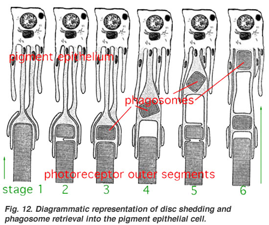
The energy requirements of the retina are even greater than that of the brain. This is due to the additional energy needed to continuously renew the photoreceptor outer segments, of which 10% are shed daily. Energy demands are greatest during dark adaptation when its sensitivity is most enhanced. The choroid supplies about 75% of these nutrients to the retina and the retinal vasculature only 25%. When light strikes 11-cis-retinal (in the disks in the rods and cones), 11-cis-retinal changes to all-trans-retinal which then triggers changes in the opsins. Now, the outer segments do not regenerate the retinal back into the cis- form once it has been changed by light. Instead the retinal is pumped out to the surrounding RPE where it is regenerated and transported back into the outer segments of the photoreceptors. This recycling function of the RPE protects the photoreceptors against photo-oxidative damage and allows the photoreceptor cells to have decades-long useful lives. Histology of the Eye, edited by William Krause, Dept. Pathology and Anatomical science, University of Missouri School of Medicine Eye, Brain, and Vision – online book – by David Hubel Kolb, H., Fernandez, E., & Nelson, R. (2003). Webvision: The neural organization of the vertebrate retina. Retinal layers image. NeuroScience 2nd Ed at United States National Library of Medicine Jeremy Nathans's Seminars: "The Vertebrate Retina: Structure, Function, and Evolution" Retina – Cell Centered Database
Text derived from https://en.wikipedia.org/w/index.php?oldid=1378897214 under CC BY-SA 4.0; modifications have been made.